Lab Practice – Observing the
heart - Are ventricles different?
The circulation of the blood in humans is
double and complete.
Objective
To check if there are anatomical differences in the heart between its right side and its left side.
Hypothesis
If there are two different circulatory systems, maybe there are anatomical differences in the heart.
To check if there are anatomical differences in the heart between its right side and its left side.
Hypothesis
If there are two different circulatory systems, maybe there are anatomical differences in the heart.
Do you think there
will be any differences? Choose a hypothesis and justify it.
A)
The left ventricle will be thicker and more resilient.
B)
The right ventricle will be thicker and more resilient.
C)
There is not significant anatomical difference.
Materials
tweezers, scalpel, hand needle,scissors, dissecting
tray and probe
Procedure – Part 1
To do this
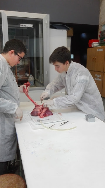
practice, each group has brought a real heart to the laboratory. Some have come
with a lamb's heart; others have come with a pig heart. Both of them are
very similar to the human heart, so somehow we could say that we have done a
dissection of... "our own heart"!
First, we
have observed the external shape of the heart and its relationship with the
lungs. Some volunteers have investigated the ventilation capacity of the lungs
of the poor lamb, or pig. Very daring!
We have
learned to distinguish between the front and rear areas of the heart. Then, we have
identified the right side of the heart and the left side, and the corresponding
chambers (atria and ventricles).
On its
surface, you can clearly see the coronary arteries and veins, which are
responsible for nurturing and maintaining this vital organ healthy and running.
The task of
identifying the veins that carry blood to the heart has been a little more
complicated. The arteries that leave the ventricles, which are responsible for
pumping blood to all the body's cells, have been easier to identify than veins.
But in the end we managed to recognize all the major veins and heart-related
arteries.
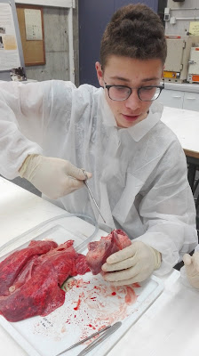
Then, we
proceeded to dissect the heart to better understand its internal structure.
First we have opened it through the pulmonary artery, and so we have seen the right
side of the heart (right atrium and right ventricle) and the valves that let
the blood through the heart (tricuspid
valve) and then through the pulmonary artery to the lungs. These heart
valves allow blood to pass only in one direction, preventing the fluid from returning back.
Subsequently, we have opened the left side of the heart with scissors
cutting through the aorta. We have seen the valve between the left
atrium and left ventricle (mitral valve) quite well. We have also seen the flap
valves, which are just where the aorta connects to the ventricle.
Definitely,
the internal structure of the heart is a real show!
And we have found that, among the
three hypothesis we had set ourselves at the beginning, the right one is the first:
the
left ventricle is thicker and stronger than the right. That is because
the left side of the heart is responsible for pumping blood throughout the
body, through greater or general circuit, while the right side of the heart is
only responsible for operating the pulmonary circuit.


